1. Resolution
SE resolution: 1.2nm or less @ 30kV
3.0nm or less @ 1kV
2. Magnification: 18 – 1,000,000X or more
(Based on 800x600 pixel monitor display)
3. System Operation: Microsoft® Windows XP or more
Standard Mouse, Knob-set and Keyboard Operation
4. Electron optics
1) Electron gun: ZrO/W Schottky emission electron gun
2) Accelerating Voltage: 0.5 – 30kV or more (should be adjustable in 100V steps)
3) Landing voltage: 0.1k – 2kV or more (should be adjustable in 100V steps)
4) Beam current: 200nA maximum or more
5) Lens System : Three-stage electromagnetic lens reduction system
6) Astigmatism: Electromagnetic 8-pole X,Y method correcting unit
7) Scanning coil: Two-stage electromagnetic deflection method
8) Objective Lens Aperture: Click stop, strip aperture with five positions or more
(open, 150μm, 70μm, 30μm, and 20μm)
* Aperture Heater should be Standard.
* Position Sensor should be Standard.
9) Beam blanking: Electromagnetic type(beam is blanked when image is frozen)
10) SE Bias Accelerator Plate: Improves SE collection efficiency at low voltages and short working distances
11) Objective Lens: Super conical lens
12) Image Shift: ± 50um or better at 15mm WD
5. Electron optics alignment
Alignment mode: Beam alignment mode
Aperture alignment mode
Stigma alignment mode
Ultra low voltage alignment mode
Auto focus alignment mode
Automated optical axis alignment and astigmatism correction mode
6. Detectors: Everhart-Thornley secondary electron detector (Standard)
Top detector for high resolution imaging through-the-Lens(TTL) or equivalent.
7. Vacuum System
1) Automatic evacuation:
Fully automatic vacuum sequence with pneumatic valves
Failsafe protection against loss of power, air pressure and vacuum
User friendly and accurate computer control of vacuum levels
2) Vacuum pumps:
Two ion-pumps (30 l/s x1, and 20l/s x1) or more
Turbo molecular pump (240 l/s) x1 or more
뿌리장비 예약
장비 상세정보
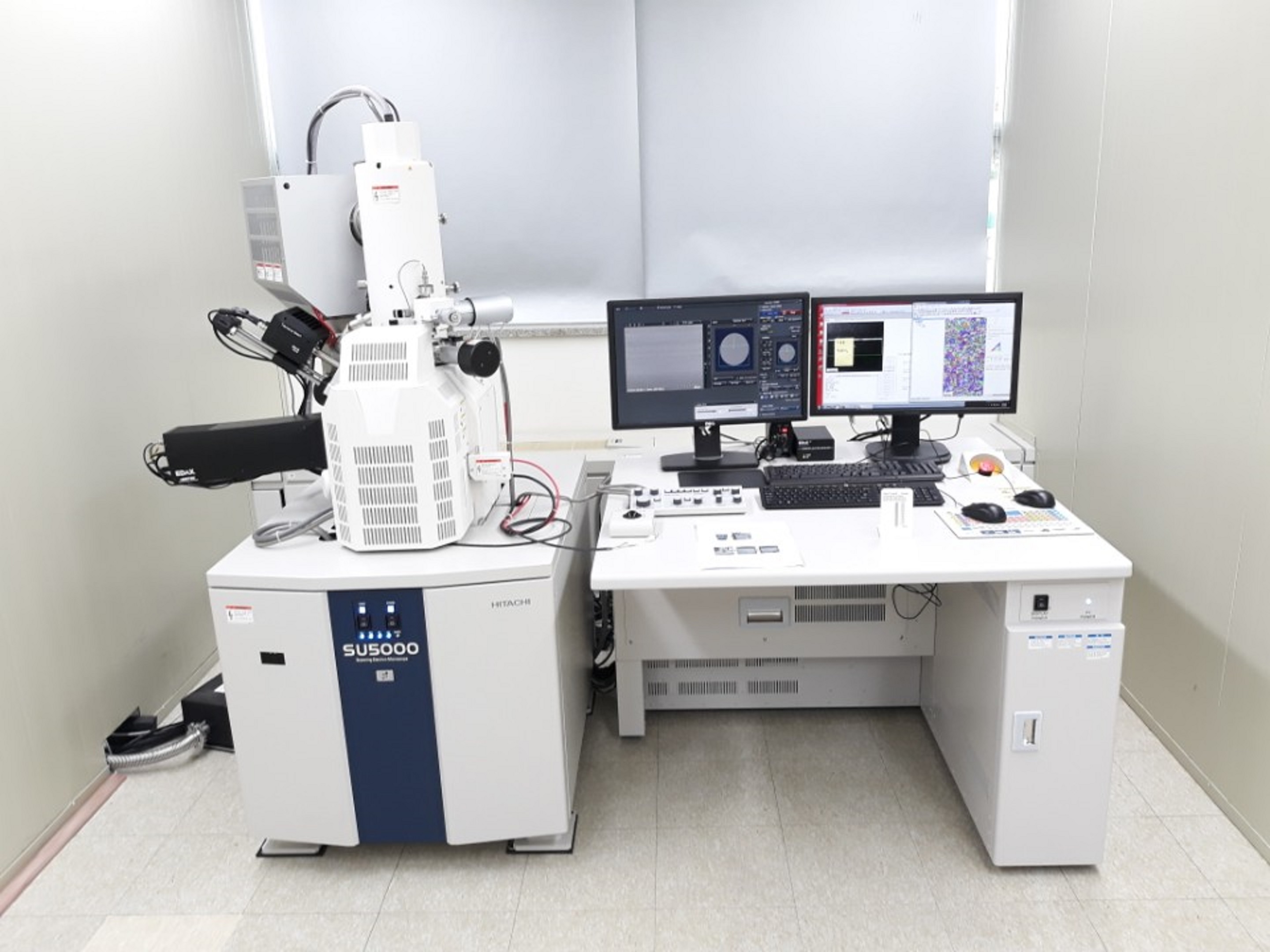
| 장비사진 | 장비명 | 주사전자현미경(Field emission scanning electron microscope) | ||
|---|---|---|---|---|
|
제작사 | HITACHI | 모델명 | SU5000 |
| NTIS | NFEC-2018-06-244260 | E-TUBE | 1805-A-0198 | |
| 장비분류 | 분석장비 | 기술분야 | 소성가공 | |
| 보유센터 | 원주뿌리기술지원센터 | 장비상태 | 정상 | |
| 구축일 | 2018-05-23 | 구축비용 | 427,976,070원 | |
| 수수료 | 60,000원/시간당 | 바우처 사용 | 사용가능 | |
| 담당자 | 김성탁 | 연락처 | ||
| 매뉴얼 | ||||
주요사항
원리 및 특징
주사전자현미경은 전자선이 시료면 위를 주사(scanning)할 때 시료에서 발생되는 여러 가지 신호 중 그 발생확률이 가장 많은 이차전자(secondary electron) 또는 반사전자(back scattered electron)를 검출하는 것으로 대상 시료를 관찰한다.
사용예
주사전자현미경에서는 주로 시료 표면의 정보를 얻을 수 있고 시료의 두께, 크기 및 준비에 크게 제한을 받지 않는다. 주사전자현미경은 광학현미경에 비해 집점심도가 2배 이상 깊고, 또한 광범위하게 집점을 맞출 수 있어 입체적인 상을 얻는 것이 가능하다.
활용분야
전계방사형 주사전자현미경은 강원권 주력산업인 자동차, 의료기기 및 신소재산업관련 기업의 기술고급화 및 R&D지원을 위해 구축예정인 장비로 고배율의 이미지 분석을 통해 미세조직을 관할할 수 있으며, 부분적인 성분과
집합조직을 함께 측정할 수 있는 분석 장비입니다.
집합조직을 함께 측정할 수 있는 분석 장비입니다.